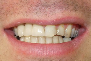
 Enamel is the external part of the Crown of the teeth. This substance, which covers the dentin, is the hardest and the most mineralized body. With dentin, cementum, and dental pulp, it is one of the four major tissues that make up the tooth. It is normally visible dental tissue, supported by an underlying layer of dentine. It consists 96% of mineral matter, the remainder being composed of water and organic matter. The mineral part is mainly composed of a network of crystals of calcium hydroxyapatite (Ca10 (PO4) 6 (OH) 2). The high percentage of minerals in enamel is responsible not only strength, but its Friability. Dentin, which is less mineralized and less brittle, is essential to support and compensate for the weaknesses of the enamel.
The color of the enamel is yellow to light gray. Since enamel is semi-translucent, the color of dentin (or any dental repair material) under enamel affects strongly the appearance of the tooth.
Enamel varies in thickness over the surface of the tooth. It is more thick at the level of the top of the dental Crown (more than 2.5 mm) and more thin on the enamel-cement (JEC) junction. Cementum and bone, the organic matrix of the enamel does not contain collagen or keratin; It has instead of glycoprotein rich in tyrosine (amélogénines, énamélines, and protein “tuft”) whose role is thought to help the growth of enamel using construction framework, among other functions. This organic matrix also contains polysaccharides.
Structure
The enamel is formed by the juxtaposition of basic structures called cords or enamel prisms. Each Prism mineralized 4 to 8 µm in diameter through the enamel, the junction to the surface of the tooth enamel-dentine.
These prisms are of hydroxyapatite crystals surrounded by a sheath of organic nature, nested one within the other. In cross-section, they resemble a lock hole, with the upper part oriented towards the Crown of the tooth and the base oriented towards the root.
The provision of the crystals within each Prism is very complex. The améloblastes (or adamantoblastes), cells which initiate enamel formation, and the extensions of Tomes influence both on the crystals. The head of the Prism enamel crystals are oriented parallel to the long axis of the latter while those base differ slightly from the long axis.
The arrangement in the space of the enamel prisms is understood more clearly that internal structure. The enamel prisms are located in a row along the tooth, and within each rank, the long axis of the Prism is generally perpendicular to the underlying dentin. In the final teeth, close the enamel-cement (JEC) junction of enamel prisms tip slightly over to the root of the tooth.
The area around the prism of enamel consists of interprismatique enamel. The latter has the same composition as enamel in Prism; It is however a histologic distinction between the two because Crystal orientation is different in each case. The limit where crystals of prismatic enamel and crystals of interprismatique enamel to touch is called prismatic sheath.
Striae of Retzius are strips that appear on enamel when it is observed in cross section under the microscope. Formed by the variation of the diameter of the extensions of volumes, these bands attest to the growth of enamel in a manner similar to the rings of a tree. The perikymaties are shallow grooves corresponding to the line formed by the Striae of Retzius on the surface of the enamel. Darker than the other bands, the neonatal line separates enamel formed before and after the birth.
Development
Enamel formation is part of the overall process of formation of a tooth. When observed the tissues of the tooth development in the microscope, can distinguish different clusters of cells, such as diamond-like body (Enamel organ), the dental lamina and the dental papilla. The generally recognized stages of the development of the tooth are the bud stage, the CAP, the stage stage Bell and stage Crown (or calcification). Enamel formation is visible only from the Crown stage.
The amélogenèse (or enamel formation) takes place after the beginning of the appearance of dentin, with cells called améloblastes. human enamel forms at a rate of about 4 µm per day, starting at the level of the future location of the cupsides of the tooth, in the 3rd or 4th month of pregnancy approximately. As in all human processes, the creation of the enamel is complex, but can generally be divided into two stages. The first step, called secretory stage, involves proteins and an organic matrix form a partially mineralized enamel. The second stage, called stage of maturation, completes enamel mineralization.
In the secretory stage, the améloblastes are polarized column-shaped cells. Enamel proteins are produced at the level of granular endoplasmic reticulum of these cells, and then released into the extracellular medium where they form what is called the matrix of the enamel. This matrix is then partially mineralized by the enzyme alkaline phosphatase. When this first layer is formed, the améloblastes away from the dentin, allowing the development of extensions of Tomes on the apical part of the cell. Enamel formation continues around the adjacent améloblastes (which induces the creation of a surface partitioned, or “sinks”, which houses the extensions of Tomes) and also around the tip of each extension of volumes (which induced the filing of a matrix of enamel in each well). The matrix within the well will become the prism of enamels term and partitions become term interprismatique enamel. The only factor of distinction between the two is the orientation of hydroxyapatite crystals.
In the phase of maturation, the améloblastes transport substances used in the formation of enamel. The most notable aspect of this phase at the tissue level is that these cells become striated, or have a wavy border. This shows that the améloblastes have changed their function: producer (cf the secretion phase), they become conveyor belts. The proteins used to the final mineralization process compose most of the transported material. The most notable proteins involved are amélogénines, améloblastines, enamélines and “proteins tuft”. During this process, the amélogénines and améloblastines are removed after use, but the énamélines and “proteins tuft” are left in the enamel. At the end of this phase, the mineralization of the enamel is completed.
After the phase of maturation, but before the tooth to appear in the mouth, the améloblastes decompose. Enamel, unlike most other tissues of the body, therefore has no way to renew itself. After a destruction of enamel by action of bacteria or injury, neither the body nor the dentist will not repair the fabric of the enamel. In addition, enamel may be affected by non-pathological processes. Staining of teeth over time can result from exposure to substances such as tobacco, coffee and tea, but the color of the tooth can also gradually darken with age. Indeed, the darkening is in part due to materials that accumulate on the level of the enamel, but is also one of the effects of the underlying dentin sclerotization. In addition, the enamel becomes with age less permeable to fluids, less soluble in acid, and contains less water.
Destruction
Dental caries
The high content mineral of enamel, which makes this tissue the hardest of all human tissue, it also likely to suffer a demineralization process that often occurs as dental caries. Demineralization can occur for several reasons, but the main cause of the development of caries is the ingestion of sugars.
The sugar candy, sugary drinks and even fruit juices play an important role in tooth decay and therefore the destruction of enamel. The mouth contains a large number and a wide variety of bacteria, and when sucrose, the most common of sugars, covers the surface of the tooth, some oral bacteria interact with it to form lactic acid, which decreases the pH in the mouth. Enamel hydroxyapatite crystals are then demineralized, allowing a greater bacterial invasion and more in depth in the tooth. The bacteria the most involved in tooth decay is Streptococcus mutans, but the number and the species of bacteria vary depending on the progression of dental destruction.
In addition, the morphology of the tooth is the most common location for a beginning of dental caries is located in notches, holes and cracks in the enamel. This is not surprising because these locations are very difficult or impossible to reach with a toothbrush tooth and allow bacteria to settle there. When demineralization of enamel occurs, a dentist may use a sharp instrument, such as a dental hook, and feel that “this addiction” to the location of the decay. As enamel demineralizes is always and is unable to prevent the bacteria from clinging, underlying dentin becomes interference. When the dentin, which supports the enamel in normal times, is destroyed by decay or another health concern, the enamel is unable to supplement its friability and detaches easily from the tooth.
Cariogénicité (ability to cause dental caries) of a food dependent factors, how for example the duration during which the sugars remain in the mouth. It is not the amount of sugar ingested, but the frequency of ingestion of sugar which is the main factor responsible for caries. When the pH in the mouth decreased intake of sugar, enamel demineralizes is and remains vulnerable for 30 minutes approximately. Eat more sugar at once does not increase the time of demineralization. Similarly, eat less sugar in a single decision-making does not decrease the duration of demineralization. Thus, eat a large quantity of sugar only once in the day is less harmful than a very small amount ingested many times throughout the day. For example, in terms of oral health, it is best to eat a single dessert to dinner to a bag of candy snack throughout the day.
Bruxism
In addition to bacterial invasion, enamel is subject to other destructive forces. Bruxism (compulsive grinding of tooth) destroys enamel very quickly. The rate of wear of enamel, called attrition, is 8 µm per year in normal times. It is a common mistake to believe that enamel wears mainly by chewing. In fact, the teeth rarely touch during chewing. Normal contact of teeth is compensated physiologically by the tooth ligament and the arrangement of the teeth when the mouth is closed. Really destructive forces are the movements of parafonction (such as suction it digitale (most often the thumb) or an object (nipple or linen), or bruxism), which can cause irreversible damage enamel.
Other causes of destruction of enamel
Other processes of non-bactérienne enamel destruction include abrasion (by foreign elements such as brushes tooth, or pins required pipe pipes between the teeth), erosion (by chemical processes involving acids, for example the action of lemon juice or gastric juice when it goes back the esophagus), and sometimes the abfraction (by forces of compression or tension).
Oral hygiene and fluoride
Cleaning teeth
The enamel is therefore very vulnerable to demineralization, and following the ingestion of sugar attacks are daily. Thus, dental health is essentially preventive methods to reduce the presence of food debris and bacteria in contact with enamel. Is used for this in most of the countries the tooth brush, which reduces the number of bacteria and food particles on enamel. A few isolated societies do not have access to such equipment but use of other objects such as pieces of wood, to clean their teeth. Can be used to clean the surface of the enamel between two adjacent teeth, dental wire. However, neither the tooth brush, or dental thread cannot enter the hollow microsopiques of enamel, but good general oral hygiene habits can in General prevent sufficient development of the bacterial population to prevent the onset of dental caries.
Email and fluorine
Fluoride is found naturally in water, but at rates highly variable. It is also present in all foods of marine origin (fish, seafood, salt sea etc.). The rate of fluorine in drinking water is 1 ppm (part per million). Fluoride helps the prevention of caries by binding to enamel hydroxyapatite crystals, which makes this last more resistant to demineralization, and therefore more resistant to the development of caries. However, an excess of fluorine can be problematic by causing disturbances called dental fluorosis. Fluorosis is overexposure to fluoride, especially between 6 months and 5 years, and is manifested by the appearance of stains on teeth. The appearance of the teeth becomes for the less unsightly, even though the incidence of caries on this kind of enamel is very low. To avoid this problem, can use filters in the areas where the rate of fluorine in tap water is too high to reduce it. The rate of fluorine is considered toxic when it exceeds 0.05 mg of fluoride per kg of body mass. Fluorine in toothpaste pasta or baths of mouth do seem to have limited both effect the fluorosis on the prevention of caries. It appears that only fluoride ingested in the fluoridated salt or water can have a real action, be it positive or negative. only the surface of the enamel is reached by the pulp fluorine toothpaste.
Saliva
Saliva has a protective role on enamel. It contains several elements protective, regulators, acting individually or organizing in real systems of defence against bacteria, but also by providing ions necessary to re-mineralization of the tooth, when it is not too damaged.
Effect of dental techniques
Dental repair
Numerous dental repairs require to remove at least part of the enamel. Generally, the purpose of this practice is to access the infected, such as dentin or the dental pulp underlying layers, for example in the case of a dentistry, an Endodontics or the installation of a Crown. The enamel may also have disappeared before any appearance of decay (see # Destruction).
Acid etch
Invented in 1955, this technique uses a dental teeth. It is commonly used in dentistry. By dissolving minerals in enamel, the teeth removes 10 µm of the surface of the enamel and involves the creation of a porous layer of 5 to 50 µm in depth. This makes speckled enamel at the microscopic level and increase the adhesion of the materials used for dental repair (dental amalgam, composite dental etc.).
The effects of the bonding vary depending on the duration of its application, the type of teeth used, and State of the enamel on the etch is applied. We also think that the results would vary depending on the orientation of the crystals of enamel.
Tooth whitening
Note: they also found the term “bleaching” despite its passive ordinary meaning (bleaching of hair).
Incoming search terms:
- any dentist using mineralization techniques


