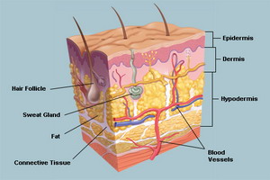
 Whether in washing, shaking hands or the grasping of objects: our skin is daily in use, and must work to it. But how is the largest organ precisely constructed and what features does the skin for the human body?
The skin serves not only as a protective sheath of the body, but it also regulates the body temperature and water resources. With its over four million receptors, it conveys us cold, heat or pain. Depending on the size and weight, the skin on average weighs 3.5 kg. Its surface measures 1.5 to 2 square meters.
Together with the so-called skin attachment formations such as hair, nails, sweat, perfume and sebaceous glands, characterises the unique appearance of human skin and also meets a variety of functions:
Functions of the skin at a glance
Protection: The skin protects from the body’s interior from external influences.
Social structure and reproduction: By which constrict blood vessels of the skin or can extend and liquid through the skin are delivered (sweating), body temperature can be regulated.
Water balance: The skin protects the body from losing too much fluid and at the same time can be a specific fluid loss to (for example, by making fluid and salts).
Meaning: The skin is our greatest sensory organ, because she perceives as warmth or cold, touch or pain.
Immune system: The skin many cells of the immune system, such as mast cells, Langerhans cells and T-cells found in.
Exchange of information by body signals: expression of an emotional response the flush or setting, might that arises through an expansion or constriction of skin blood vessels, especially in the area of face, neck and décolleté.
The skin consists of several layers, which can be roughly split into the skin (epidermis), DermIS (DermIS) and subcutaneous tissue (hypodermis). Each of the three layers of the skin performs its own function. So, the subcutaneous tissue serves mainly as a fat storage, while the epidermis is a protective function with their horny layer. The DermIS is responsible with their special structures for the sense of touch. The epidermis and the upper DermIS accommodate special cells of the immune system, the T-cells.
Skin (epidermis)
The thick visible skin (epidermis) is distributed across the body different dick – the strongest bodies located at the soles of the feet and the clutching. There, the epidermis is up to two millimeters thick.
The top layer of the skin is composed of brick-like layered, scale-shaped skin cells without nucleus. It is dead keratinocytes (horn-forming cells) are in deeper cell layers, that then migrate to the surface. Meanwhile they change their shape and their cell contents steadily, until they are finally becoming more and more verhornen and be repelled as Horn scales. This is an ongoing process, so that the epidermis is constantly renewed by scaling and replication.
The epidermis is divided from inside to outside in five layers:
Basalzellschicht (stratum basal): in the Basalzellschicht is the formation of keratinocytes stem cells by cell division. From the Basalzellschicht, a new layer is continuously over several stages of regeneration. Melanocytes and Merkel cells are embedded in the Basalzellschicht. The melanocytes are pigment, which is responsible for the coloring and tanning of the skin: the melanin. Merkel cells are associated with nerve fibers and convey a part of touch. In palms and soles, they occur more frequently than in other body areas.
Stachelzellschicht (stratum spinosum): in the Stachelzellschicht, the keratinocytes are connected mesh-like detention zones (ultrastructural). In this layer can occur in skin diseases to water retention and thus to the formation of bubbles. Here you find also Langerhans cells, which are part of the immune system of the skin.
Körnerzellschicht (stratum granulosum): the keratinocytes in the Körnerzellschicht contain increasingly so-called Keratohyalinkörner that bring about the progressive keratinisation.
Highly refractive layer, gloss layer (stratum lucidum): the stratum lucidum is found only on the thicker parts of the epidermis, e.g. on the palms and soles. This layer is very narrow, cell borders or cell nuclei are no longer to recognize.
Horny layer (the stratum corneum): clump the horny cells emerged from the keratinocytes with Horn substances of the skin in the stratum corneum. The Horn scales are then repelled. Until the cells reach from the bottom of the layer to the surface and are repelled about two weeks pass. The stratum corneum doesn’t hardly through water and water soluble substances. Low molecular substances can however penetrate them. Special lipids (Ceramides) that prevent that dry out the skin along with the horny cells are a major component of this skin barrier. In addition, they protect against the ingress of contaminants or irritants and pathogens in the body. The barrier function is also supported by a hydrolipid film of sweat and sebum covering the surface of the skin. This skin barrier is weakened when the skin very long comes into contact with water.
DermIS (DermIS)
The DermIS (DermIS), which consists of a dense network of elastic and Collagen fibers located between the upper and subcutaneous tissue. This network gives the skin strength and elasticity. Other components of the DermIS are blood and lymph vessels, immune defence cells, hair follicles, nerves and skin glands. In addition, high-level pressure receptors mediate the sense of touch in the dermis. This so-called Meissner Corpuscle are well represented, especially in the sensitive fingertips.
The DermIS is divided into two layers:
Papillary layer (stratum papillare): the papillary layer is located directly on the epidermis and zapfenartig protruding into it. Many small blood vessels (capillaries), as well as melanocytes are found in the papillary layer. But also special cells of the immune system (mast cells) occur here in large numbers.
Network layer (stratum reticulare): the network layer consists mainly of firmer collagen fiber bundles. It follows on the papillary layer and borders directly on the skin.
Subcutaneous tissue (Subcutis)
The subcutaneous tissue (Subcutis) consists of loose bouquets and adipose tissue and forms the link between the skin and the underlying structures. The subcutaneous fatty tissue includes a fixed number of fat cells, which include different-sized Fetttropfen depending on the nutritional status. The fat is used as energy storage and heat insulator. The subcutaneous tissue of the skin also allows the displacement. It comes to pathological accumulation of water (edema), is the fluid accumulation in this layer.
In addition, more structures such as nerves, blood vessels, are hair follicles, glands, smooth muscle cells and nerve cells that perceive vibrations (father Pacini lamellar bodies) in the subcutaneous tissue.
Skin appendage
Epidermal appendage are training skin. The skin attachment formations include hair, nails, sweat, perfume and sebaceous glands.
Hair (Pili) almost invariably occur all over the body. Only palms, soles, lips, and parts of the sex organs are saved by the hair growth. Distinction between Terminal hair and Wollhaaren. The Terminal hair includes scalp hair, eyelashes, eyebrows, pubic hair, armpit hair, beard and chest hair (in men), nose hair and hair in the outer ear canal – they are usually longer, stronger and pigmented.
The remaining body hair, which is usually quite short, colorless and sparsely fails in comparison to the Terminal hair is one of the Wollhaaren.
Nails (Ungues) protect fingers and toes. Finger- and toenails consist of about 0.5 mm thick nail plate (Horn plate), which is located in the nail bed.
Fingernails grow about three times as fast as toenails: per week approximately 0.5 to 1.5 millimeters formed fingernail newly. Until a fingernail is fully grown after, it takes about three to four months.
The skin attachment formations include the glands of the skin, like Sebaceous, welding, and scent glands in addition to hair and nails. Sebaceous glands are found mainly in hairy skin areas. The tallow they give off, ensures that the hair and skin are supple and gives the hair shine. At the same time, is tallow part of the acid mantle and thus serves to protect of the skin. At heat the sebaceous glands produce which is why many people increasingly to fight have in wintertime with dry skin usually more sebum or on cold of less.
Draining of sebum from the sebaceous glands, known as blackheads can form. If in addition the sebaceous glands bacteria settle, inflammation and finally an acne may arise.
Sweat glands can be found, but above all in the area of the forehead, palms, and the soles of the feet in almost all areas of the skin. However, the lips and the foreskin of the penis are free of sweat glands. A total two to four million sweat glands in the skin are approximately. They form the salty sweat, which serves to the one the temperature regulation of the body and on the other hand contribute to the acid mantle of the skin.
Scent glands are present mainly in the armpit, in the genital and anal area, and women in the areola of the nipple. You are controlled by sex hormones and begin only with the beginning of puberty, to work.


